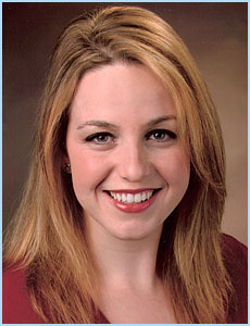

Dr. Miriam Van Allen was born and raised in Los Angeles, California. She graduated Cum Laude with a Bachelor of Science degree in Psychobiology from the University of California Los Angeles in 2000. After completing her undergraduate degree, Dr. Van Allen then attended the UCLA School of Dentistry, where she was president of the Alpha Omega dental organization, president of the student chapter of the American Association of Women Dentists, and vice-president of her dental school class. She received her Doctor of Dental Surgery degree in 2005.
After dental school, Dr. Van Allen moved to
 Colorado to complete her orthodontic specialty residency at the University of Colorado, Denver. During her orthodontic residency training, she received an advanced certification in Invisalign clear braces treatment and attended numerous lectures on orthodontic miniscrew implants, along with completing a research project involving digital modeling for orthodontic treatment plans. Dr. Van Allen enjoys enhancing her orthodontic knowledge by pursuing the most advanced proven techniques the orthodontic profession has to offer. In return, Dr. Van Allen is able to provide her patients with only the best of care in a compassionate manner.
Currently, Dr. Van Allen enjoys being back in Southern California, taking full advantage of both the sun and surf. She is a member of the American Association of Orthodontics, the Los Angeles Dental Society, the California Dental Association, and the American Dental Association. In addition to her involvment in both the local and dental communities, she loves the theater, dancing, traveling, and spending time with her family and friends.
We are proud to have her as a part of our team.